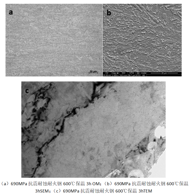
金相样品,先在预磨机上进行预磨,然后在150、320、600和1000号砂纸上依次水磨后,在抛光机上进行机械抛光,抛光后的试样置于4 ml 硝酸(HNO3) + 96 ml 无水乙醇(CH3CH2OH)混合溶液浸蚀10~20 s,在Olympus GX53光学显微镜中观察其金相组织。600℃ 3h后微观组织没有明显变化。SEM样品,先在预磨机上进行预磨,然后在150、320、600和1000号砂纸上依次水磨,在抛光机上进行机械抛光后,置于4 ml HNO3 + 96 ml CH3CH2OH混合溶液浸蚀15~25 s,采用FEI Quanta650热场发射扫描电子显微镜分析,观察贝氏体形貌、尺寸与分布等。MA岛组元发生了分解。TEM样品,从待分析试样的相应部位线切割厚度约为0.3 mm的薄片,水磨减薄至50 μm以下,在-20~-15℃,采用6 ml 高氯酸(HClO4)+94 ml CH3CH2OH的双喷液对薄片进行双喷,双喷电流为50 mA。双喷后的试样使用FEI Tecnia G 20透射电子显微镜(附带能谱分析仪EDS)进行观察。在基体中析出大量的析出相。
The metallographic samples were pre-ground on a pre-grinding machine, then water-ground on sandpaper no. 150, 320, 600 and 1000 successively, and then mechanically polished on a polishing machine. After polishing, the samples were immersed in a mixed solution of 4 mL nitric acid (HNO3) + 96 mL anhydrous ethanol (CH3CH2OH) for 10~20 s. The metallographic structure was observed by Olympus GX53 optical microscope. After 600℃ for 3h, the microstructure did not change significantly. SEM samples were pre-ground on a pre-grinding machine, and then waterground on sand paper 150, 320, 600 and 1000 in turn. After mechanical polishing on a polishing machine, they were immersed in 4 mL HNO3 + 96 mL CH3CH2OH mixed solution for 15-25 s. FEI Quanta650 thermal field emission scanning electron microscope was used to observe the shape, size and distribution of Baines. The composition of MA Island has been decomposed. TEM samples were prepared by wire cutting 0.3mm thin slices from the corresponding parts of the sample to be analyzed. The thin slices were reduced to less than 50 μm by water milling. At -20~-15℃, the thin slices were double-sprayed with 6 mL perchloric acid (HClO4) +94 mL CH3CH2OH with a double injection current of 50 mA. FEI Tecnia G 20 transmission electron microscope (EDS) was used to observe the samples after double spraying. A large number of precipitated phases are precipitated in the matrix.