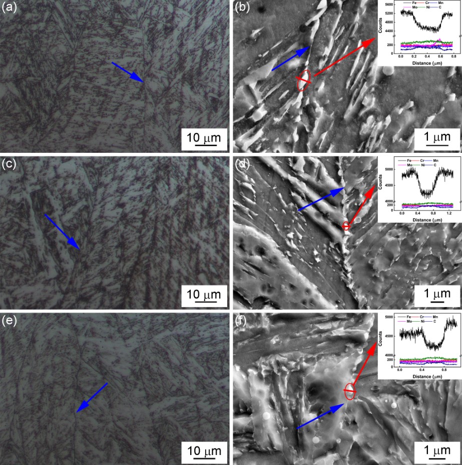
焊缝金属的基体主要由典型的贝氏体形态组成,并且可以看到原始奥氏体晶界(蓝色箭头的位置)。在这些显微照片((a)、(c)和(e))中可以清楚地观察到许多焊接金属枝晶。具有较暗对比度和较亮区域的轮廓分别被认为是枝晶间和枝晶干区域。精细的微观结构在更高的放大倍数下通过 SEM 进一步表征。在原奥氏体晶界上可以清楚地观察到大量析出相。这些析出相的组成分布通过 SEM-EDS 线扫描表征,析出相的线分布在(b)、(d)和(f)中显示,表明析出相可能是 M3C,其中 M 主要是铁。比较所有三种焊缝金属的整体显微组织,没有观察到明显的差异,证实 V 含量的轻微变化对基体结构没有明显影响,并且在原始奥氏体晶界上有大量析出相。
The matrix of the weld metals predominantly consisted of the typical morphology of bainite, and the prior austenite grain boundaries were visible (positions of blue arrows). Numerous weld metal dendrites could be clearly observed within these micrographs ((a), (c), and (e)). The outlines with darker contrast and lighter regions were considered as the inter-dendritic and dendrite core regions, respectively. The fine microstructures were further characterised by SEM at higher magnifications. Numerous precipitates could be clearly observed on the prior austenite grain boundaries. The composition distribution of these precipitates was characterised by SEM–EDS line scans, and the line profiles across the precipitates are presented in (b), (d), and (f), revealing that the precipitates could be M3C, where M was mainly Fe. Comparing the overall microstructures of all three weld metals, no obvious differences could be observed, confirming that the slight change in the V content had no appreciable influence on the matrix structure and large precipitates on the prior austenite grain boundaries.