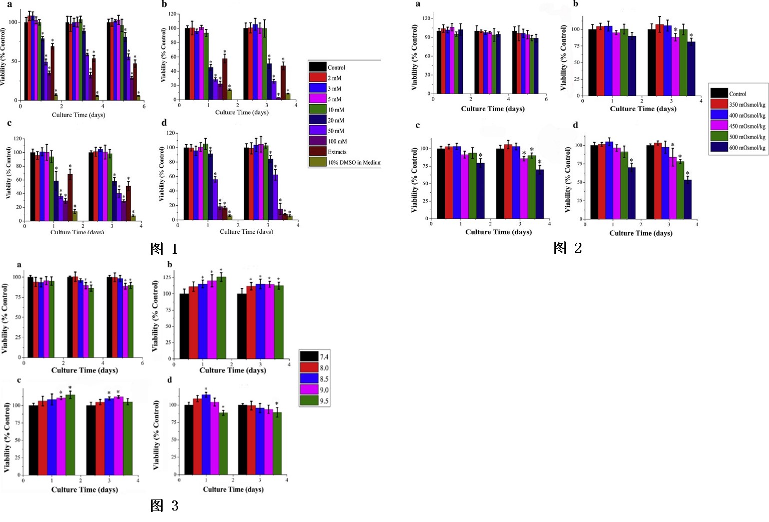
使用一系列具有上升镁离子浓度的培养基来测试成骨细胞、MC3T3-E1、BMSC和L929的镁剂量耐受性(图1)。 根据现行ISO第5部分标准,细胞存活率高于75%可认为对医疗器械无毒性风险,因此我们将细胞存活率为75%的Mg离子浓度定义为安全水平。 绘制了在一定范围的镁离子浓度下孵育 72 小时(最常用的时间点)的四种细胞类型的细胞活力,以获得最大耐受剂量的镁离子(细胞活力高于 75%)。 对于 L929 和成骨细胞,安全水平为 35 mM,而对于 BMSCs 和 MC3T3-E1,安全水平仅为 15 mM。培养基中的渗透压随着 Mg 离子浓度的增加而升高,因此应用一系列具有用 5 M 氯化钠调整的上升渗透压值的培养基来测试细胞对高细胞外渗透压的反应。 虽然培养基渗透压的增加可以诱导对细胞增殖的抑制作用,但是当培养基中的渗透压值从 300 mOsmol/kg 增加到 500 mOsmol/kg 与提取物中的渗透压值匹配时,细胞活力仅降低 10%(图2)。细胞对碱性环境的敏感性可能在很大程度上取决于细胞类型的选择。当选择L929和成骨细胞进行细胞毒性试验时,较高的pH值(超过8.5)对细胞活力有不利影响(约下降10%),而碱性环境模拟了BMSCs和MC3T3-E1的细胞生长,特别是在早期 (图3)。
A series of culture media with ascending Mg ion concentrations were used to test the Mg dose tolerance of osteoblasts, MC3T3-E1, BMSC and L929 (Fig. 1). According to the current ISO standards of Part 5, cell viability higher than 75% could be considered with no toxic risks for medical devices, so we defined the Mg ion concentration with 75% cell viability as the safety level. The cell viability of the four cell types incubated for 72 h (the most commonly used time point) in a range of Mg ion concentrations was plotted to get the most tolerated dose of Mg ions (cell viability above 75%). For L929 and osteoblasts, the safety level was 35 mM, while for BMSCs and MC3T3-E1, the safety level was only 15 mM. Osmolality in media raised with increasing Mg ion concentrations, so a series of media with ascending osmolality values adjusted with 5 M sodium chloride were applied to test cell responses to high extracellular osmolality. Although an increase in osmolality of the medium could induce inhibitory effects on cell proliferation, only 10% cell viability was reduced when osmolality values in medium increased from 300 mOsmol/kg to 500 mOsmol/kg matching with the osmolality values in the extracts (Fig.2). Sensitivity of cells to alkaline environments might be greatly dependent on cell types selected. Higher pH (over 8.5) showed detrimental effect on cell viability (approximately 10% decrease) when L929 and osteoblasts were chosen for cytotoxicity tests, while the alkaline environment simulated cell growth of BMSCs and MC3T3-E1, especially at the early time point (Fig. 3).