
国家材料腐蚀与防护科学数据中心
National Materials Corrosion and Protection Scientific Data Center
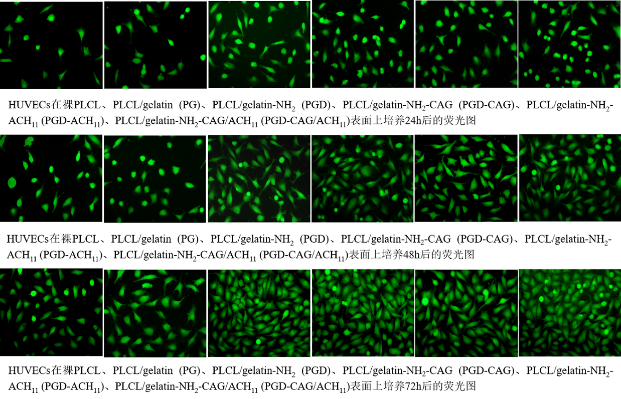
样品表面HUVEC的数量均随培养时间的增加而增加。在培养1天后,PLCL和PG表面上仅观察到少量HUVECs,并且它们表现出椭圆形且细胞未充分铺展。然而,PGD表面的细胞粘附数量明显较高且增殖迅速,在培养3天后几乎覆盖了PGD整个表面,明显高于其它所有组。这可能归因于仿生PDA涂层能够有效促进细胞的粘附和增殖。此外,在三个时间点,PGD-CAG表面HUVECs粘附量均高于PGD表面。这主要得益于CAG肽能够通过表面的受体整合素选择性靶向内皮细胞,导致HUVECs在材料表面的粘附能力大大增强。
The number of HUVECs increased with culture time on all samples in the fluorescence images. It was found that a few HUVECs were observed on the PLCL and PG surface after 1 day of culture and they exhibited oval-shaped with inefficient cell spreading. However, the cell adhesion on the PGD surface was significantly more than on the PG and PLCL surfaces after 1 day of culture. Meanwhile, the HUVECs proliferated rapidly and covered almost the entire surface of the PGD surface at 3 days. The results confirmed that development of PDA coating on adhesion and proliferation of cells. Furthermore, it could be seen that there was a higher HUVECs amount on the PGD-CAG surface compared with that on the PGD surface at all three time points. It was mainly because CAG peptides can interact with the receptors on the HUVECs membrane, thus resulting in further enhanced cell adhesion.
 国家材料腐蚀与防护科学数据中心 |
 国家高能物理科学数据中心 |
 国家基因组科学数据中心 |
 国家微生物科学数据中心 |
 国家空间科学数据中心 |
 国家天文科学数据中心 |
 国家对地观测科学数据中心 |
 国家极地科学数据中心 |
 国家青藏高原科学数据中心 |
 国家生态科学数据中心 |
 国家冰川冻土沙漠科学数据中心 |
 国家计量科学数据中心 |
 国家地球系统科学数据中心 |
 国家人口健康科学数据中心 |
 国家基础学科公共科学数据中心 |
 国家农业科学数据中心 |
 国家林业和草原科学数据中心 |
 国家气象科学数据中心 |
 国家地震科学数据中心 |
 国家海洋科学数据中心 |