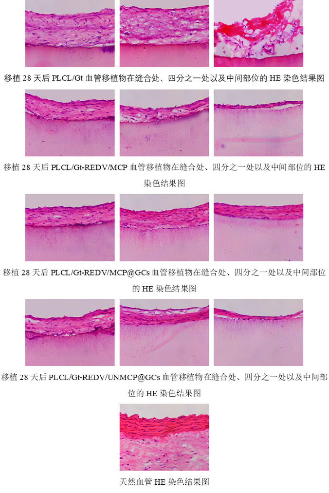
PLCL/Gt移植物的缝合部位、四分之一处和中间部分被较厚的新内膜覆盖,并导致凝血基质沉积。此外,PLCL/Gt-REDV/MCP和PLCL/Gt-REDV/UNMCP@GCs移植物的这些部位也形成了新内膜,但其厚度从缝合线到中间部分逐渐减小,表明新内膜没有完全形成,尤其是在中间站点。 对于PLCL/Gt-REDV/MCP@GCs血管移植物,新生内膜完全形成,厚度均匀,结构致密,与天然血管一致。
The suture site, quarter and mid portions of PLCL/Gt grafts were covered by significantly thick neointima, and resulted in coagulation matrix deposition. Besides, these sites of PLCL/Gt-REDV/MCP and PLCL/Gt-REDV/UNMCP@GCs grafts also formed neointima, but its thickness gradually decreased from suture to middle portion, indicating that the neointima didn’t completely form, especially in middle site. For PLCL/Gt-REDV/MCP@GCs vascular graft, the neointima completely formed with uniform thickness and dense structure, which was consistent with nature blood vessels.