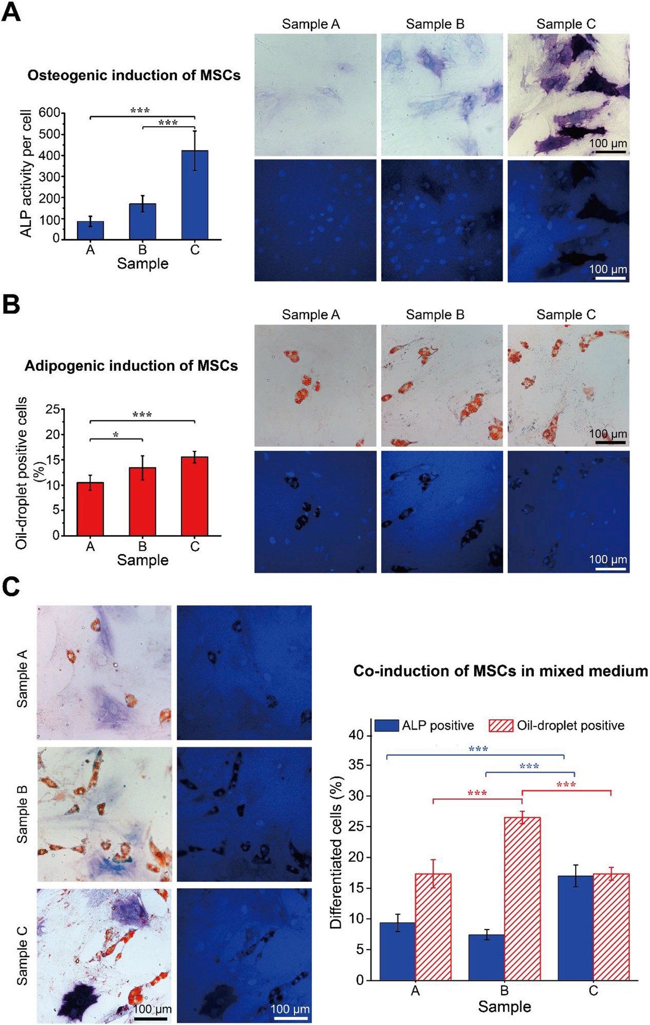
测定和统计了细胞在三种不同降解速率的纳米图案化的PEG基水凝胶(C>B>A)表面的分化结果。(A)骨髓基质干细胞在材料表面进行7天成骨分化诱导后的分化比例统计结果。细胞进行碱性磷酸酶(ALP)染色和细胞核染色,上方为ALP染色的明场显微图,下方为核染色的荧光显微图。(B)骨髓基质干细胞在材料表面进行7天成脂分化诱导后的分化比例统计结果。细胞进行油红染色和细胞核染色,上方为油红染色的明场显微图,下方为核染色的荧光显微图。(C)骨髓基质干细胞在材料表面进行7天成骨/ 成脂共分化诱导后的分化比例统计结果。细胞进行ALP染色、油红染色和细胞核染色,左侧为ALP、油红染色的明场显微图,右侧为核染色的荧光显微图。
Bright-field images and statistics of MSC differentiation on samples at different degradation stages. (A) Osteogenesis of MSCs after osteogenic induction for 7 d. Cells experienced ALP staining and nucleus staining prior to the optical microscopic observations. (B) Adipogenesis of MSCs after adipogenic induction for 7 d. Cells were submitted to oil-droplet staining and nucleus staining prior to observations. (C) Co-induction of MSCs in a mixed induction medium for 7 days. Cells experienced ALP staining, oil-droplet staining and nucleus staining prior to the microscopic observations.