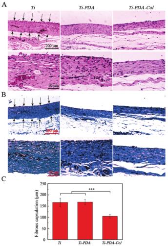
钛(Ti),多巴胺钛(Ti-PDA),及多巴胺和胶原修饰的钛(Ti-PDA-Col)植入SD大鼠30天后,通过(A)H&E染色和(B)Masson三色染色对样品周围的纤维包囊进行组织学分析。图中箭头指示的是纤维包囊的边界。(C)来自所示样品的纤维包囊厚度的统计结果。数据表示为均值±SD(n = 3),并通过单因素方差分析法进行分析。当p < 0.001时,显著性差异用三个星号标记。
Histological analysis of the biopsy of the capsule surrounding indicated samples by (A) H&E staining and (B) Masson’s trichrome staining after 30 days of implantation in SD rats. The arrows indicate the boundary of fibrous capsulation. (C) Statistical results of the thickness of fibrous capsulation from the indicated samples. Data are presented as mean ± SD (n = 3) and analyzed by one-way ANOVA. Significant differences are marked with three asterisks when p < 0.001.