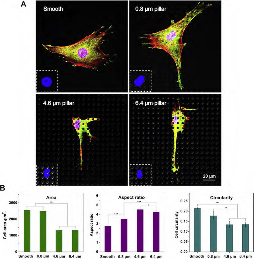
测定了细胞在0.8, 4.6 以及 6.4 um高度PLGA微柱,以及光滑表面上的细胞行为。(A)细胞荧光显微图。细胞核采用DAPI染色,显示为蓝色;细胞骨架微丝F-actin用phalloidin-TRITC染色,显示红色;vinculin蛋白用anti-vinculin单克隆抗体及荧光二抗染色,显示绿色。(B)细胞铺展面积、长径比、以及圆度。
Behaviors of cells on micropillar arrays with pillar heights of 0.8, 4.6 and 6.4 um as well as smooth films were examined. Representative fluorescence micrographs of MSCs after 24 h of culture are presented in Fig A. The corresponding adhesion parameters such as projected area, AR and cell circularity were also quantified, with the results shown in Fig B.