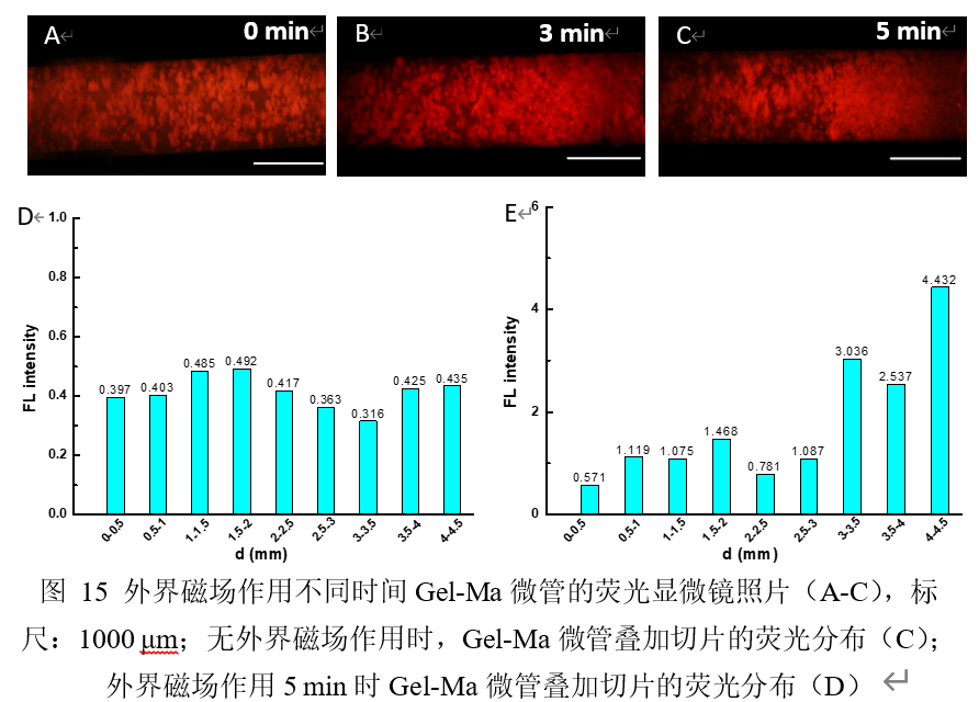
图A-C为外界磁场作用不同时间后,Gel-Ma微管内部GIMs形成的梯度分布形貌图,红色荧光为交联GIMs发出的荧光。荧光强度梯度随着磁场作用时间越长越明显,即可形成具有药物梯度的微管。为了进一步探究微管内部的药物梯度的形成,选用模型药物BSA-FITC,将具有BSA-FITC梯度的微管用冰冻切片的方法切成100 μm的厚度,然后酶标仪读取BSA-FITC的荧光值,图D和E显示无梯度的微管在0-4.5 mm的距离中的荧光强度较接近,无明显的规律;而具有梯度的微管在0-4.5 mm的距离中荧光强度呈递增趋势,表明了BSA-FITC在微管内部呈梯度分布,可以反向推出GIMs在外界磁场的作用下呈梯度分布,从微管的一端到另外一端,浓度逐渐增加。
Figures A-C show the gradient distribution morphology of GIMs inside Gel-Ma microtubules after an external magnetic field is applied for different periods of time. The red fluorescence is the fluorescence emitted by the cross-linked GIMs. The fluorescence intensity gradient becomes more obvious as the magnetic field acts for a longer time, and a microtube with a drug gradient can be formed. In order to further explore the formation of the drug gradient inside the microtubules, the model drug BSA-FITC was selected, and the microtubules with the BSA-FITC gradient were cut into a thickness of 100 μm by the method of cryosectioning, and then the fluorescence of BSA-FITC was read by a microplate reader Figures D and E show that the fluorescence intensity of microtubes with no gradient is closer in the distance of 0-4.5 mm, and there is no obvious law; while the fluorescence intensity of microtubes with gradient is increasing in the distance of 0-4.5 mm. , Which shows that BSA-FITC presents a gradient distribution inside the microtube, and it can be inferred that GIMs present a gradient distribution under the action of an external magnetic field, and the concentration gradually increases from one end of the microtube to the other end.