
国家材料腐蚀与防护科学数据中心
National Materials Corrosion and Protection Scientific Data Center
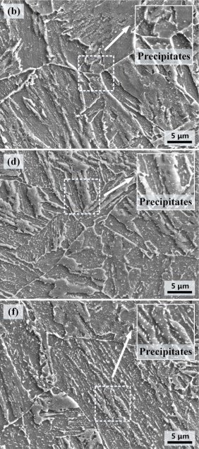
用扫描电子显微镜(SEM)对微观结构进行了表征,试样分别取自平板的亚表面、t/4和t/2位置。采用机械研磨、抛光、4% nital溶液腐蚀的制备SEM纵向横截面试样,使用Zeiss Ultra-55扫描电子显微镜金相显微镜进行显微组织观察。
The microstructure was characterized by scanning electron microscope. Samples were taken from the subsurface, T/4 and T/2 positions of the plate respectively. The SEM longitudinal cross section samples were prepared by mechanical grinding, polishing and etching with 4% nital solution, and the microstructure was observed by Zeiss Ultra-55 scanning electron microscope and metallographic microscope.
 国家材料腐蚀与防护科学数据中心 |
 国家高能物理科学数据中心 |
 国家基因组科学数据中心 |
 国家微生物科学数据中心 |
 国家空间科学数据中心 |
 国家天文科学数据中心 |
 国家对地观测科学数据中心 |
 国家极地科学数据中心 |
 国家青藏高原科学数据中心 |
 国家生态科学数据中心 |
 国家冰川冻土沙漠科学数据中心 |
 国家计量科学数据中心 |
 国家地球系统科学数据中心 |
 国家人口健康科学数据中心 |
 国家基础学科公共科学数据中心 |
 国家农业科学数据中心 |
 国家林业和草原科学数据中心 |
 国家气象科学数据中心 |
 国家地震科学数据中心 |
 国家海洋科学数据中心 |