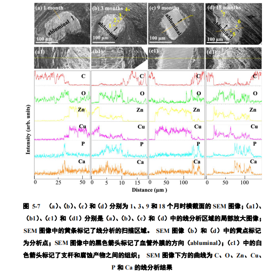
图 5-7 显示了在 1、3、9 和 18 个月时间点所选横截面的微观形貌和元素线分析结果。在每个时间点的 SEM 图像中,均可以识别出三个具有代表性区域:一种是 Zn-Cu 支架杆,在该区域仅发现 Zn 和 Cu 信号;一种是在 Zn-Cu支杆周围观察到的腐蚀产物,具有显著的 Zn,Cu,O,Ca 和 P 元素富集;第三种是组织,显示出明显的 C 信号,没有 Zn 和 Cu 峰。图 5-7(c1)中的白色箭头标记了降解的支杆被正常组织所取代的位置,该区域具有明显的组织信号特点。图 5-7(d)显示了一个完全降解的 Zn-Cu支架杆残留区域,在该区域内,只有腐蚀产物而没有发现锌铜合金的金属残留。在支架杆和腐蚀产物区都可以观察到 Zn 和 Cu 峰的共现(图 5-7),这表明这两种元素是共扩散的。
Figure 5-7 shows the micro-morphology and element line analysis results of the selected cross-sections at 1, 3, 9 and 18 months. In the SEM image at each time point, three representative regions can be identified: one is the Zn-Cu support rod, in which only Zn and Cu signals are found; the other is around the Zn-Cu support rod The observed corrosion products have significant enrichment of Zn, Cu, O, Ca and P elements; the third type is the structure, which shows a clear C signal without Zn and Cu peaks. The white arrow in Figure 5-7 (c1) marks the position where the degraded strut is replaced by normal tissue. This area has obvious tissue signal characteristics. Figure 5-7(d) shows a completely degraded Zn-Cu stent rod residue area. In this area, there are only corrosion products and no metal residues of zinc-copper alloy. The co-occurrence of Zn and Cu peaks can be observed in the stent rod and corrosion product area (Figure 5-7), which indicates that these two elements are co-diffused.