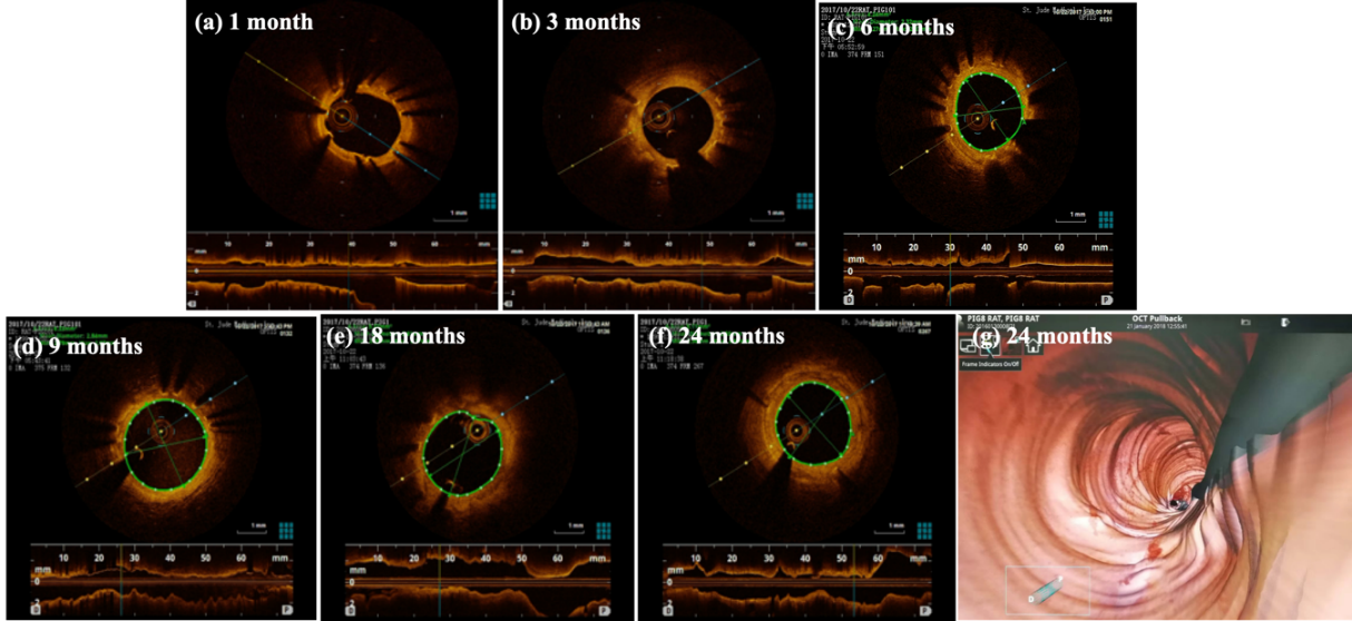
图5-5为Zn-Cu支架植入后,各个随访时间点的OCT图。
(a)-(f):在第1、3、6、9、18和24个月的时间点植入Zn-Cu支架的血管段的截面图像。周围分布的亮点是支架撑杆,后面带有黑色阴影;支柱内部是新生的内膜;(g):在24个月的时间点植入Zn-Cu支架的新生血管膜的新生内膜的3D图像(顶视图)
血管壁的结构包括内膜、中膜和外膜,支架植入后发生内皮化以及内膜重建,是血管损伤修复的重要指标[31,32]。在OCT图像上,内层黄色高亮反光的环状部分为血管内膜;图(a)-(d)中围绕内膜、明显可见的亮点为支架杆,支架杆后侧跟有黑色放射状阴影。图5-5(a)显示,锌铜支架植入1个月后,血管内壁已经形成完整的新生内膜,这意味着植入后1个月内内皮化过程已经完成。
在图5-5(a)-(c)中,OCT图像上的支架杆清晰可见;从第9个月开始(图5-5(d)),支架杆变模糊;到本研究的终点(24个月),OCT图像中的支架杆几乎变得不可见(图5-5(f))。支架杆在OCT图像上由清晰到模糊的变化过程,反应了Zn-Cu支架的冠脉血管内的逐步降解。
从Zn-Cu支架植入1个月直至研究结束,OCT图像上的新生内膜都是光滑的,且未观察到血栓形成的迹象。说明锌铜支架在动物体内没有引发严重的内膜增生或者血栓。
Figure 5-5 shows the OCT diagram of each follow-up time point after Zn Cu stent implantation.
(a) - (f): cross sectional images of vascular segments implanted with Zn Cu stents at the time points of 1, 3, 6, 9, 18 and 24 months. The bright spots distributed around are support rods with black shadows behind; The inner part of the pillar is the neointima; (g) : 3D image of neointima of neovascular membrane implanted with Zn Cu stent at 24 months (top view)
The structure of vascular wall includes intima, media and adventitia. Endothelialization and intimal reconstruction after stent implantation are important indicators of vascular injury repair [31,32]. On OCT images, the annular part with yellow highlight and reflection in the inner layer is the intima of blood vessels; In figures (a) - (d), the obvious bright spot around the intima is the stent rod, and there is a black radial shadow on the back of the stent rod. Figure 5-5 (a) shows that a complete neointima has been formed on the inner wall of the blood vessel one month after the implantation of the zinc copper stent, which means that the endocortization process has been completed within one month after the implantation.
In figures 5-5 (a) - (c), the support rod on the OCT image is clearly visible; From the 9th month (Fig. 5-5 (d)), the support rod becomes blurred; By the end of this study (24 months), the stent rod in the OCT image became almost invisible (Fig. 5-5 (f)). The change process of stent rod from clear to fuzzy on OCT image reflects the gradual degradation of Zn Cu stent in coronary artery.
From 1 month after Zn Cu stent implantation to the end of the study, the neointima on OCT images was smooth, and no signs of thrombosis were observed. It shows that zinc copper stent does not cause serious intimal hyperplasia or thrombosis in animals.