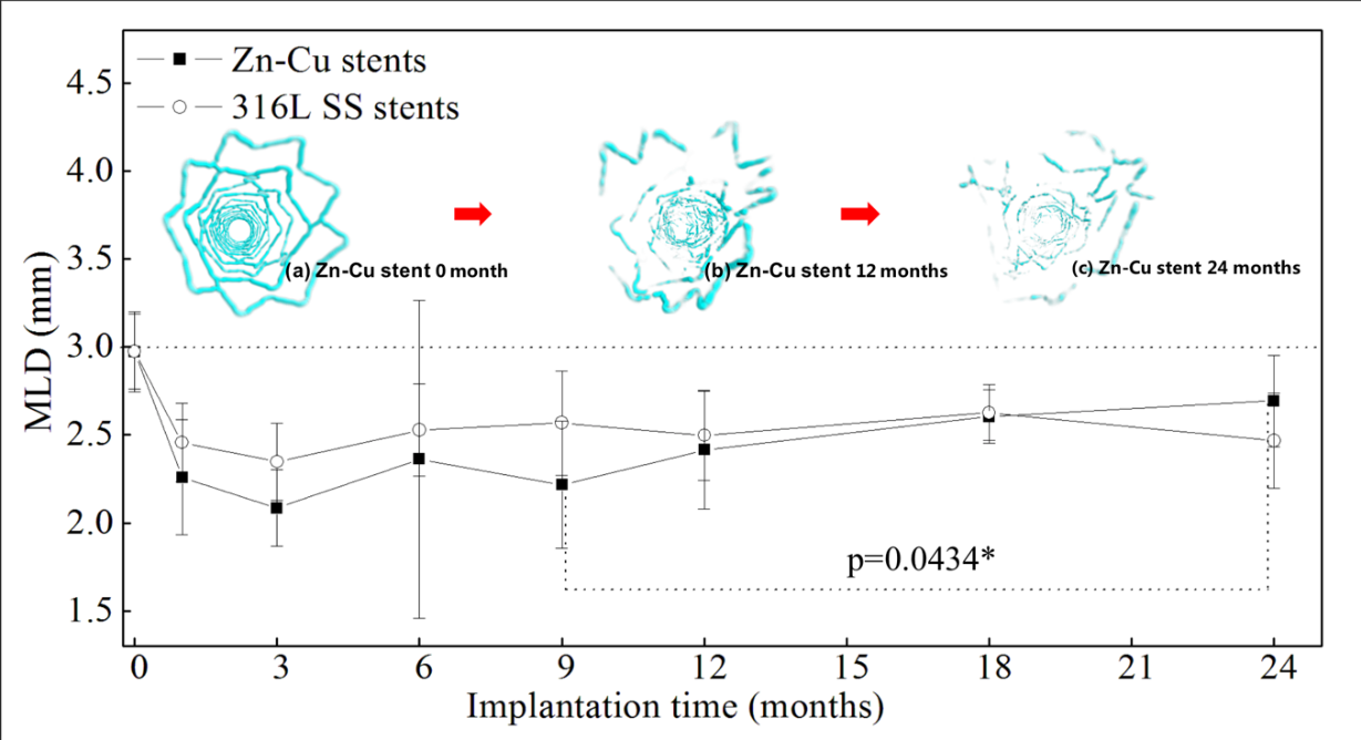
图 5-4 Zn-Cu组和316L SS组各时间点(n=5或6)的MLD
插图为:(a)植入前的Zn-Cu支架,(b)12个月后的Zn-Cu支架残留物,(c)24个月后的Zn-Cu支架残留物的Micro-CT三维图像(俯视图);符号“*”表示Zn-Cu组9个月和24个月的MLD值差异有统计学意义(P<0.05)
由表5-2可知道,Zn-Cu支架和316L SS支架在植入动脉前后靶血管直径基线相当(参考血管直径p=0.43;瞬时血管直径p=0.50)。植入段血管的最小管腔直径(MLD)变化可以直接反映两组器械支架内再狭窄的发展情况。
表5-2和图5-4显示了在每个时间点通过定量冠状动脉造影(QCA)和LLL评估的MLD结果(n=5或6)。图5-4 中的插图(a)、(b)和(c)是通过Micro-CT三维重建得到的0、12和24个月时间点上Zn-Cu支架残留物的3D图像(俯视图)。结果表明,在24个月的研究期间,Zn-0.8Cu组和316L SS组的MLD和LLL结果均无显著差异。提示锌铜支架的机械性能与316L SS支架相当。
图5-4可见,Zn-Cu支架植入段段血管的最小管腔直径MLD在9个月以后呈现上升趋势。到本次试验的终点(24个月),MLD值已经和9个月时出现了显著性差异的(p=0.43),表明由于Zn-Cu支架的降解,猪冠脉血管发生了正向重构,管腔直径开始变大。在镁合金冠脉支架和可降解聚合物支架的动物试验研究中,均发现过类似现象[26,30]。
表5-2和图5-4所示的LLL和MLD结果表明,锌铜支架克服了先前报道的可降解支架的局限性(包括较低的径向强度和降解过程中产生的炎症引发的严重管腔丢失等),并且随着降解进程,支架对血管的束缚作用也得以解除。
Figure 5-4 MLD of Zn Cu group and 316L SS group at each time point (n = 5 or 6)
The illustrations are: (a) Zn Cu stent before implantation, (b) Zn Cu stent residue after 12 months, (c) micro CT three-dimensional image of Zn Cu stent residue after 24 months (top view); The symbol "*" indicates that there is significant difference in MLD values between Zn Cu group at 9 months and 24 months (P < 0.05)
It can be seen from table 5-2 that Zn Cu stent and 316L SS stent have the same target vessel diameter baseline before and after arterial implantation (reference vessel diameter P = 0.43; instantaneous vessel diameter P = 0.50). The change of minimum lumen diameter (MLD) of implanted vessels can directly reflect the development of in stent restenosis in the two groups.
Table 5-2 and figure 5-4 show the MLD results (n = 5 or 6) assessed by quantitative coronary angiography (QCA) and LLL at each time point. Illustrations (a), (b) and (c) in Figure 5-4 are 3D images (top view) of Zn Cu scaffold residues at 0, 12 and 24 months time points obtained by micro CT 3D reconstruction. The results showed that there was no significant difference in MLD and LLL results between zn-0.8cu group and 316L SS group during the 24 month study period. It is suggested that the mechanical properties of zinc copper stent are equivalent to that of 316L SS stent.
Figure 5-4 shows that the minimum lumen diameter MLD of the implanted segment of Zn Cu stent shows an upward trend after 9 months. By the end point of this trial (24 months), there was a significant difference between MLD value and that at 9 months (P = 0.43), indicating that due to the degradation of Zn Cu stent, porcine coronary vessels underwent positive remodeling and the lumen diameter began to increase. Similar phenomena have been found in animal experimental studies of magnesium alloy coronary stents and degradable polymer stents [26,30].
The LLL and MLD results shown in table 5-2 and figure 5-4 show that zinc copper stents overcome the limitations of previously reported degradable stents (including low radial strength and severe lumen loss caused by inflammation during degradation), and the binding effect of stents on blood vessels can be relieved with the degradation process.