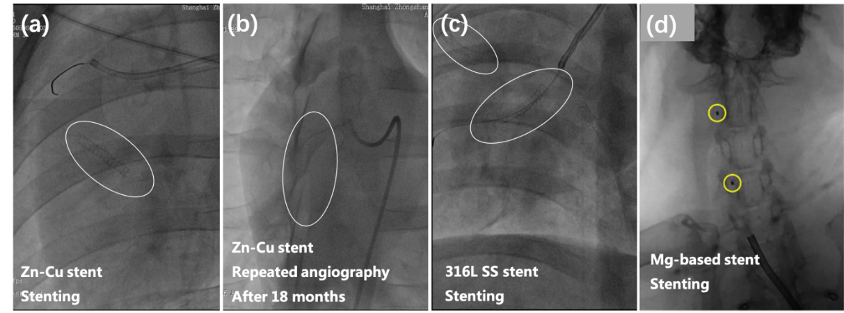
图 5-3 支架的X射线可探测性:(a)植入Zn-Cu支架的猪冠状动脉术中造影(白圈突出显示支架位置);(b)植入Zn-Cu支架的猪冠状动脉术后18个月造影(白圈突出显示支架位置);(c)植入316L SS支架的猪冠状动脉术中造影(白圈突出显示支架位置);(d)植入镁基支架的新西兰白兔颈总动脉术中造影[23](球囊上的不透射线标记用黄色圆圈突出显示,以帮助定位支架)
材料X射线可探测性主要取决于其材料密度及电子密度的高低。锌的电子密度高于镁、铁或者高分子聚合物,因此在可降解支架中具有优势。图5-3(a)显示了手术过程中猪冠状动脉内的Zn-Cu支架:锌铜支架在X光下显影清晰,支架杆扩张充分,支架充盈后形态理想。对比图5-3(c)可见,锌铜合金支架在X光下的显影性与传统支架(316L SS支架)相当,说明其能够满足临床操作对显影性的要求。相比之下,在5-3(d)镁支架血管造影图像中几乎看不到支架形貌。镁支架的定位依赖于球囊上的不透射线标记。这些标记物与不透射线的碘化造影剂一起,也可以实现支架在动脉内的精确放置。然而,这种显影方法无法满足支架植入后进行后扩张调整的要求[24]。
图5-3(b)为植入18个月后,Zn-Cu支架植入段血管在X光下的图像:此时从在X光下已经难以发现Zn-Cu支架的图像,说明Zn-Cu支架发生了降解、导致密度下降。
Figure 5-3 X-ray detectability of stent: (a) intraoperative angiography of porcine coronary artery implanted with Zn Cu stent (white circle highlights the position of stent); (b) Angiography 18 months after implantation of Zn Cu stent in porcine coronary artery (white circle highlights the position of stent); (c) Intraoperative coronary angiography of pigs implanted with 316L SS stent (white circle highlights the position of the stent); (d) Intraoperative angiography of common carotid artery in New Zealand white rabbits implanted with magnesium based stent [23] (the radiopaque mark on the balloon is highlighted with a yellow circle to help locate the stent)
The X-ray detectability of materials mainly depends on their material density and electron density. The electron density of zinc is higher than that of magnesium, iron or polymer, so it has advantages in degradable scaffolds. Figure 5-3 (a) shows the Zn Cu stent in porcine coronary artery during the operation: the zinc copper stent is clearly developed under X-ray, the stent rod is fully expanded, and the shape is ideal after stent filling. Compared with FIG. 5-3 (c), the development of zinc copper alloy stent under X-ray is equivalent to that of traditional stent (316L SS stent), indicating that it can meet the development requirements of clinical operation. In contrast, almost no stent morphology was seen in 5-3 (d) magnesium stent angiography images. The positioning of magnesium stent depends on the radiopaque mark on the balloon. These markers, together with radiopaque iodized contrast agents, can also achieve the precise placement of stents in the artery. However, this development method cannot meet the requirements of post expansion adjustment after stent implantation [24].
Figure 5-3 (b) shows the X-ray image of the blood vessels in the implanted section of Zn Cu stent 18 months after implantation: at this time, it is difficult to find the Zn Cu stent under X-ray, indicating that the Zn Cu stent has been degraded and the density has decreased.